
How to easily understand sinus rhythm abnormalities in dogs and cats
Let me show you this:
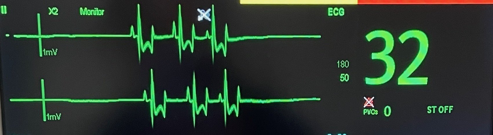
This is the (at the time, terrifying) trace belonging to a patient I anaesthetised not long ago.
They were in for investigations into severe, chronic gastrointestinal disease. I was monitoring them for an ultrasound and endoscopy, and they were bradycardic, with long pauses between heartbeats.
So I popped them on an ECG (like we do for all of our anaesthetised patients), and… yep. Sinus arrest.
So if you were in this situation, how would you react? Would you understand how to recognise this rhythm, and know how to manage the patient?
Well, if you want to learn more about rhythms like this, you’re in luck - because today I’m sharing the 7 sinus rhythm abnormalities we see in dogs and cats. You’ll leave this post with a better understanding of what each of these is, how to spot it, and which need treatment.
And that’s not all…
If you want more information on ECGs, I’m hosting an event inside the Medical Nursing Academy very soon that you’ll NOT want to miss!
Introducing… ‘Name That Arrhythmia!’
An interactive webinar where we’ll use a few simple questions to break down different ECG traces, understand more about the different arrhythmias we see, and understand how to treat and nurse these patients.
So if you want to feel more confident interpreting ECGs and managing arrhythmia patients, you need to be at this event!
And - at the time of this post going live - the doors to the medical nursing academy are officially OPEN!
So, head to www.medicalnursingacademy.com, grab your spot, and start learning inside with me and the rest of the members inside our community!
Want more information on the academy and whether it’s the right place for you? Drop me a DM on Instagram and let’s chat!
So let’s talk sinus arrhythmias…
Several arrhythmias affect the sinus (aka sinoatrial) node within the heart.
As a reminder, our sinoatrial node acts as our pacemaker, spontaneously generating impulses that spread throughout the atria, causing contraction, before heading to the atrioventricular node, and ultimately causing ventricular contraction. This, in turn, causes the ejection of blood from the heart.
This is our normal sinus rhythm - where we have a P wave, followed by our QRS complex, and T wave. It is a regular rhythm, and the rate at which the SA node fires to create our normal sinus rhythm varies - both depending on the species, and on the situation (e.g. rest, exercise, and stress).
But what about when our patient’s sinus rhythm isn’t normal? Well, today we’re going to look at the following abnormal rhythms:
Sinus bradycardia
Sinus tachycardia
Sinus arrhythmia
Sinoatrial block
Sinus arrest
Atrial standstill
Sick sinus syndrome
Sinus Bradycardia
Sinus bradycardia is a regular sinus rhythm - but, as the name suggests, it’s slow.
Specifically, it is defined as a rhythm that is slower than expected for the species, and for the situation the patient is in.
For example:
Let’s say you have a blocked bladder cat present. Their heart rate is 124 beats/minute.
Is that a normal heart rate? It could be, in a chilled-out, comfortable cat at rest…
But what about a painful, stressed cat who has been admitted as an emergency?
We’d be expecting their heart rate to be sitting at around 160+, most likely. So this patient, despite having a seemingly normal heart rate, is bradycardic.
What causes sinus bradycardia?
We often see this rhythm in patients who have received anaesthetic agents, as these cause dose-dependent cardiovascular depression.
We can also see sinus bradycardia in patients with
Increased vagal tone due to certain medications or diseases like respiratory, neurological, gastrointestinal, urinary or ocular disease)
Decreased sympathetic nervous system tone due to the use of medications like xylazine, or beta-blockers
Hypothermia
Hypothyroidism
Sick sinus syndrome
So should we treat these patients?
Not usually, unless your patient has clinical signs associated with their bradycardia - like hypotension (think about your anaesthetised patients here), weakness, collapse, or exercise intolerance.
If you do need to treat them, it’s usually done by giving them an anticholinergic like atropine.
Sinus Tachycardia
Sinus tachycardia is a rapid sinus rhythm. It’s defined as a regular sinus rhythm, faster than normal, BUT appropriate for the situation that the patient is in.
If your patient is at rest, not stressed, and they have an inappropriately high heart rate - e.g. over 200 beats/minute - we should be looking for another cause of the tachycardia. These include abnormalities such as atrial or ventricular tachycardia.
So when do we see sinus tachycardia?
There are LOTS of causes of sinus tachycardia, including:
Stress
Exercise
Hyperthyroidism
Pyrexia
Pain
Hypovolaemia
Heart failure
Administration of catecholamines
Pheochromocytoma
Cardiac tamponade (e.g. secondary to pericardial effusion)
And what about treatment?
Treatment is mostly aimed at correcting the underlying cause - such as giving analgesia to a painful patient. In severe cases, we may consider giving a beta-blocker, such as atenolol.
Sinus Arrhythmia
Sinus arrhythmias are seen where there is an irregular discharge of the SA node, usually associated with the patient’s respiratory cycle. The frequency of the SA node activating/discharging varies, and usually, we see the SA speed up during respiration and decrease during pauses between breaths.
In dogs, sinus arrhythmia is a completely normal finding. It is abnormal in cats within the hospital, but it can be noted more commonly (and normally) in cats in the normal home environment.
What else causes sinus arrhythmia?
Sinus arrhythmia can also be seen in dogs due to increases in vagal tone, rather than respiration.
We can also see sinus arrhythmia in patients with wandering pacemaker - where there are taller P waves during higher heart rates, and shorter P waves during lower heart rates.
So what do we do about it?
If it is a normal finding, or not causing a problem with the patient’s blood pressure/perfusion, it does not need to be managed - only monitored.
Decreasing vagal tone - e.g. due to things like excitement and exercise, or administration of drugs to counteract it such as atropine - can resolve it.
Sinoatrial Block
SA block is a common arrhythmia in dogs - but it’s often hard to diagnose.
We see SA block when the impulse from the sinoatrial node does not get conducted through the surrounding tissues to the atria and ventricles.
This means your patient has no P wave, no QRS complex, and, instead, they have an increase in their P-P interval (ie. the interval from the P wave of one complex to the P wave of the next complex) - because the heart has ‘skipped a beat’.
Specifically, the P-P interval is exactly double what it usually is when there is SA block. However, because sinus arrhythmia is so common in dogs, it makes SA block very tricky to diagnose. Why? Because there are variations in the patient’s P-P interval with that arrhythmia, making it challenging to spot SA block.
Sinus Arrest
Sinus arrest or sinus pause is an absence of P waves for longer than with SA block - so a P-P interval of above 2 x normal.
When do we see sinus arrest?
Sinus arrest is usually caused by high vagal tone, sinus node disease, or a combination of both. We tend to see it in patients with sick sinus syndrome (which we’ll come on to in a minute!) or a very marked sinus arrhythmia.
How do we treat sinus arrest?
Where sinus arrest is causing marked bradycardia and hypotension, it is managed by administering drugs such as atropine (which is what we did in our anaesthetised patient at the beginning of this post!)
Atrial Standstill
Our next sinus node abnormality is atrial standstill. We see this very commonly in hyperkalaemic patients, as well as patients with certain cardiac diseases.
Atrial standstill is defined as the complete absence of P waves on an ECG. It occurs when the atria cannot be depolarized by the electrical impulse discharging from the SA node.
Why does this happen?
It usually occurs because:
The myocardium in the atria are unable to be depolarized, which is usually the case with hyperkalaemia
The myocardium has been damaged/destroyed by a cardiomyopathy or myocarditis, causing persistent atrial standstill.
What happens when a patient gets atrial standstill?
When there is persistent atrial standstill, the SA node can also be affected by the underlying cardiac disease. This means that our patient develops a heart rhythm controlled by the AV node (as the SA node would act as a pacemaker where it is functioning normally). This is known as a junctional escape rhythm, and it usually has a lower heart rate of around 40-65 beats/minute.
How do we manage atrial standstill?
Well, it depends on the underlying mechanism behind it. In patients with hyperkalaemia, we need to reduce that by:
Giving appropriate fluid therapy
Administering glucose
+/- Administering neutral insulin
We can also give calcium gluconate, which protects the heart - but does not drop the potassium level itself.
In a patient with persistent atrial standstill, we manage their underlying cardiac disease +/- consider pacemaker implantation where needed.
Sick Sinus Syndrome
Sick sinus syndrome is a combination of abnormalities, including sinus arrest, junctional or ventricular escape rhythms, and (possibly) supraventricular tachycardia.
These patients are usually bradycardic (tachycardia with sick sinus syndrome is rare) and present with signs such as exercise intolerance, weakness and/or syncope.
What causes sick sinus syndrome?
SSS is an acquired condition usually diagnosed in older dogs. There are certain breeds at an increased risk of the condition, including West Highland White Terriers, Cocker Spaniels and Miniature Schnauzers.
It can be caused by high vagal tone or is due to abnormalities in the SA node, surrounding tissue, or both. It can also be caused by a combination of these abnormalities AND having a high vagal tone!
We can also see AV block alongside sick sinus syndrome, because conduction abnormalities can be noted in other areas, such as the AV node.
What signs do we see in a sick sinus syndrome patient?
We see similar signs to other sinus arrhythmias, including:
Weakness
Exercise intolerance
Collapse
Syncope
Also… we need to think about looking for this disease in our at-risk patients.
So say you have a Westie present for a dental. They’re ok in themselves, but perhaps a little quiet - which the client had associated with their age.
Yes, it could be - but they could also have sick sinus syndrome - so have a good listen to these patients when they present for anaesthetics or sedations!
I like to run ours around the corridor for a little while, to see if the exercise brings their heart rate up - if they don’t have SSS, it should!
How do we treat sick sinus syndrome?
Medications to increase heart rate, like terbutaline or extended-release theophylline can be used - but these can be effective only for a short time. In some cases, affected patients will require pacemaker implantation.
So what’s the take-home message for managing these patients?
When you’re nursing patients with conduction abnormalities, always ask yourself if the heart rate is low enough (or high enough, if it’s a tachycardic patient) to be impacting their perfusion, oxygenation and blood pressure.
If it is, then you’ll be wanting to give them medications to try and adapt that heart rate and improve perfusion.
Or, do they have an underlying disease that needs to be managed, such as GI disease, hyperkalaemia, or congestive heart failure?
And if neither of those is the case, and you’ve got a patient with an acceptable heart rate and blood pressure, you may just be monitoring them. So keep an eye on their blood pressure and perfusion status, and alert the vet if anything changes.
Do you see arrhythmias like these in practice? Drop me a DM on Instagram and let me know!
And don’t forget - if you want to feel more confident identifying and managing arrhythmias, make sure you’re inside the medical nursing academy before doors close on September 1st!
References
Kittleson, M. D. 2023. Conduction abnormalities in dogs and cats [Online] MSD Veterinary Manual. Available from: https://www.msdvetmanual.com/circulatory-system/heart-disease-conduction-abnormalities-in-dogs-and-cats/heart-disease-conduction-abnormalities-in-dogs-and-cats#v76144521
Kittleson, M. D. 2023. Persistent atrial standstill in dogs and cats [Online] MSD Veterinary Manual. Available from: https://www.msdvetmanual.com/circulatory-system/various-heart-diseases-in-dogs-and-cats/persistent-atrial-standstill-in-dogs-and-cats#:~:text=being%20noted.,palliate%20clinical%20signs%20of%20CHF.
Pace, C. 2013. Common arrhythmias: the importance of ECG interpretation [Online] The Veterinary Nurse. Available from: https://www.theveterinarynurse.com/review/article/common-arrhythmias-the-importance-of-ecg-interpretation